
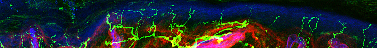
The focus of our laboratory is developing new methods to objectively diagnose, quantify and grade neuropathy of the peripheral and autonomic nervous systems. Beginning in 1990 the clinical and research activity has focused on immunostaining and confocal microscopic imaging of cutaneous nerves, principally epidermal nerve fibers (ENFs) and sudomotor nerves in skin biopsies using fluorescent labeled antibodies to Protein Gene Product 9.5 (PGP 9.5) / Ubiquitin Carboxy Hydrolase like-1 (UCHL-1), type IV collagen, Ulex and several neuropeptides. In addition we quantify mucosal nerves of the stomach, colon and ileum in gastroparesis and childhood constipation.
Confocal images of thick immunostained sections analyzed using computer software allow us to quantify the nerves fibers by number (density), length and number of branch points.
We also develop methods for improving the neurological exam. Our goal is to invent and prototype simple, portable, easy to use and understand, yet objective and highly quantitative medical devices to supplement, but not prolong, the examination. We are prototyping devices to quantify the fast conducting myelinated sensory nerves that convey tactile sensation (touch) from the finger pads, vibration and flutter (20 -40c/s) from the toes and feet, and the slow, unmyelinated sudomotor nerves to sweat glands. Our goal is to produce devices that can be easily used in a clinical setting that are fast, accurate and sensitive enough to detect small changes in the health of peripheral nerves for earlier diagnosis and detection of progression or improvement of neuropathy. Device performance is validated using the just described tissue quantification methods.